TENNIS AND GOLFER’S ELBOW
HOW MUSCULOSKELETAL ULTRASOUND IS IMPROVING
THE MANAGEMENT OF THESE COMMON CONDITIONS
By Donald Kasitinon, MD, and Reed Williams, MD – Physical Medicine and
Rehabilitation (PM&R) Sports Medicine Physicians at UT Southwestern Medical Center
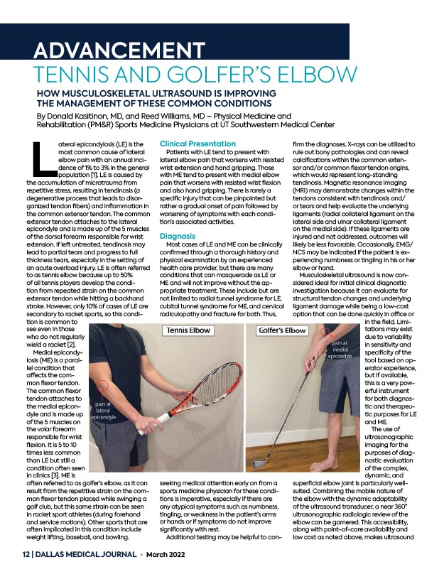
Lateral epicondylosis (LE) is the
most common cause of lateral
elbow pain with an annual incidence
of 1% to 3% in the general
population 1. LE is caused by
the accumulation of microtrauma from
repetitive stress, resulting in tendinosis (a
degenerative process that leads to disorganized
tendon fi bers) and infl ammation in
the common extensor tendon. The common
extensor tendon attaches to the lateral
epicondyle and is made up of the 5 muscles
of the dorsal forearm responsible for wrist
extension. If left untreated, tendinosis may
lead to partial tears and progress to full
thickness tears, especially in the setting of
an acute overload injury. LE is often referred
to as tennis elbow because up to 50%
of all tennis players develop the condition
from repeated strain on the common
extensor tendon while hitting a backhand
stroke. However, only 10% of cases of LE are
secondary to racket sports, so this condition
is common to
see even in those
who do not regularly
wield a racket 2.
Medial epicondylosis
(ME) is a parallel
condition that
aff ects the common
fl exor tendon.
The common fl exor
tendon attaches to
the medial epicondyle
and is made up
of the 5 muscles on
the volar forearm
responsible for wrist
fl exion. It is 5 to 10
times less common
than LE but still a
condition often seen
in clinics 3. ME is
often referred to as golfer’s elbow, as it can
result from the repetitive strain on the common
fl exor tendon placed while swinging a
golf club, but this same strain can be seen
in racket sport athletes (during forehand
and service motions). Other sports that are
often implicated in this condition include
weight lifting, baseball, and bowling.
Clinical Presentation
Patients with LE tend to present with
lateral elbow pain that worsens with resisted
wrist extension and hand gripping. Those
with ME tend to present with medial elbow
pain that worsens with resisted wrist fl exion
and also hand gripping. There is rarely a
specifi c injury that can be pinpointed but
rather a gradual onset of pain followed by
worsening of symptoms with each condition’s
associated activities.
Diagnosis
Most cases of LE and ME can be clinically
confi rmed through a thorough history and
physical examination by an experienced
health care provider, but there are many
conditions that can masquerade as LE or
ME and will not improve without the appropriate
treatment. These include but are
not limited to radial tunnel syndrome for LE,
cubital tunnel syndrome for ME, and cervical
radiculopathy and fracture for both. Thus,
seeking medical attention early on from a
sports medicine physician for these conditions
is imperative, especially if there are
any atypical symptoms such as numbness,
tingling, or weakness in the patient’s arms
or hands or if symptoms do not improve
signifi cantly with rest.
Additional testing may be helpful to confi
rm the diagnoses. X-rays can be utilized to
rule out bony pathologies and can reveal
calcifi cations within the common extensor
and/or common fl exor tendon origins,
which would represent long-standing
tendinosis. Magnetic resonance imaging
(MRI) may demonstrate changes within the
tendons consistent with tendinosis and/
or tears and help evaluate the underlying
ligaments (radial collateral ligament on the
lateral side and ulnar collateral ligament
on the medial side). If these ligaments are
injured and not addressed, outcomes will
likely be less favorable. Occasionally, EMG/
NCS may be indicated if the patient is experiencing
numbness or tingling in his or her
elbow or hand.
Musculoskeletal ultrasound is now considered
ideal for initial clinical diagnostic
investigation because it can evaluate for
structural tendon changes and underlying
ligament damage while being a low-cost
option that can be done quickly in offi ce or
in the fi eld. Limitations
may exist
due to variability
in sensitivity and
specifi city of the
tool based on operator
experience,
but if available,
this is a very powerful
instrument
for both diagnostic
and therapeutic
purposes for LE
and ME.
The use of
ultrasonographic
imaging for the
purposes of diagnostic
evaluation
of the complex,
dynamic, and
superfi cial elbow joint is particularly wellsuited.
Combining the mobile nature of
the elbow with the dynamic adaptability
of the ultrasound transducer, a near 360°
ultrasonographic radiologic review of the
elbow can be garnered. This accessibility,
along with point-of-care availability and
low cost as noted above, makes ultrasound
ADVANCEMENT
12 | DALLAS MEDICAL JOURNAL • March 2022